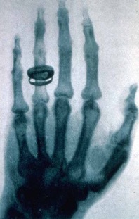
 X-ray imaging is what most people think of when they think about X-rays and is routinely used in dental and medical applications. X-rays are light with very high energy and can penetrate deeply into materials. X-rays are attenuated by matter mostly by the photo-electric effect. That attenuation depends very strongly on the X-ray’s energy and the density of electrons in the material. That means X-rays tend to pass through light elements (say, C, O, N) very easily, are partially attenuated by moderately heavy elements (say, Ca), and will be mostly stopped by very heavy elements (say, Au, Pb). This is why the dentist puts a Pb-bib over you for a dental X-ray, why the safety enclosures at beamlines have Pb-lined walls, and why an X-ray image of a hand shows the Ca-rich bones – and any break in them – enhanced compared to the muscles and flesh.
Synchrotrons are not necessary for simple X-ray imaging, but they do allow very rapid X-ray imaging, exceptionally high-resolution images, and several interesting variations on standard X-ray imaging. Geoscientists use synchrotron X-ray imaging methods at SEES beamlines to measure the time-dependent 3-dimensional interior structures of materials in geomaterials such as inclusion-rich minerals, melts at high temperatures and pressures, micro-meteorites, and micro-fossils, and to measure the draining of oil-water mixtures through sand piles and sediments. In addition, multi-wavelength imaging can be used to exploit features at X-ray absorption edges to enhance the contrast of X-ray imaging for particular elements.
X-ray Radiography
As with a familiar medical X-ray, an X-ray radiograph is an image of the attenuation of X-rays by a material, which depends strongly on the density of the material. Using a synchrotron as the X-ray source allows for very fast and high-resolution X-ray radiographs, with spatial resolutions of around 1 micron, and imaging times of 1 millisecond or less. Geoscientists use radiography at SEES beamlines to measure the precise shape of samples in multi-anvil presses, thereby directly measuring the strain on these samples under load. Other radiography experiments include high-speed imaging of a solid bead moving in a melt at high temperature and pressure to measure the viscosity of the melt, or imaging the draining of a fluid through sediments or assemblages of grains of sand.
X-ray Computed Tomography
While a single X-ray radiograph depends on the total density of the material that the X-rays pass through, Computed Tomography – just like a medical CT scan – will combine radiographs at multiple angles to produce a three-dimensional image of a material and measure the density at each voxel within the sample. Unlike, a medical CT scan, where an X-ray source and detector are spun around the human sample, tomography at SEES beamlines spins the sample in the X-ray beam. Since synchrotron sources permit very spatial resolutions, CT at SEES-operated beamlines is mostly done at the microscale, imaging the interiors of precious samples such as micro-fossils, micro-meteorites, or measuring the impacts of surface tension of oil-water mixtures on filling the spaces in sediments and sand piles. In addition, by spiking different phases with heavy elements and using the jump in X-ray absorption at a core-electron level, the contrast in density of different phases can be enhanced.
X-ray Fluorescence Mapping
As discussed in the spectroscopy section, X-ray Fluorescence (XRF) can be used to measure the abundance of different elements — especially heavy elements — in a material. At a synchrotron beamline, these measurements can be done very quickly, and at high-spatial resolution with an incident X-ray beam of a few microns in size (or even as small as 100 nm). This permits XRF to be used as a scanning probe, where a sample is moved back and forth through a fixed micro-focused X-ray beam, and the XRF spectrum is measured at each pixel. This can be used to generate images of the locations of different elements within a sample.